Juvenile dermatomyocitis with calcinosis
© Marie Jones BSc, FIMI, PgCHE, FHEA, AHCS, fCMgr
Juvenile dermatomyocitis with calcinosis received an Award of Excellence in the Biomedical catagory in BioImages 2021.
Dermatomyositis is a very rare autoimmune inflammatory disease; when the onset is in childhood it is known as Juvenile dermatomyositis (JDM).
JDM affects approximately 1:3,000,000 children per year, it is twice as common in girls than boys and the cause is unknown.
The name ‘dermatomyositis’ literally means skin muscle inflammation. Symptoms include: weak, painful muscles; skin rashes; fatigue; irritability. There may also be: oedema; arthralgia; alopecia; lipodystrophy and, as in the example here, calcinosis – hard calcium lumps under the skin or muscle. The condition in adults may be associated with malignancy.
Reason for the image creation, purpose, and audience
Now aged 72 years, calcinosis present in this patient’s arms limits his mobility. Photographs were taken of this to demonstrate the calcinosis and the sarcoma (detailed in a separate image) for the patient’s healthcare record and for referral at the Multidisciplinary Team (MDT) meeting.
The patient also gave his permission for the images to be used for teaching, publication and research purposes. He was also keen for them to be used to demonstrate the clinical presentation of JDM to health professionals and to inform other patients and their families to raise awareness of this rare condition.
Equipment and Specifications
Nikon D500 APS-C DSLR (DX); Nikon 60 mm f/2.8 D AF Micro-NIKKOR macro lens. Focal length: 60 mm (DX), equivalent to 90 mm FX.
Exposure: ISO 200; 1/125th/sec; f/25.
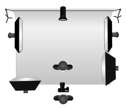
Studio lighting: Key and fill lights - x2 Bowens XMT 500 flash heads (battery powered); Rim lights - x2 Bowens 500 flash heads.
Lighting technique: Studio flash ‘cross’ lighting.
Method
I commonly use the cross-lighting technique. It is a simple but subjective method of lighting used to demonstrate texture, depth and form by accentuating the shadows in the relief areas of the image, hence, giving a more ‘3D’ effect than would normally be viewed under ‘flat’, even lighting.
Two front lights are used: one as key light and the other as fill light, together with two lights behind the subject as rim lights to ‘separate’ the subject from the background.
Challenges
The challenge when demonstrating the calcinosis in this image was to ensure that the highlights and shadows balance in order to retain detail in both. The histogram used both in camera whilst photographing and referred to in post-processing gives a useful indication as to the shadow and highlight values. I always aim to get the exposure, focus, positioning of the patient and framing correct at the time of photography to ensure integrity of the image and less digital manipulation when processing. Exposure is particularly important to ensure accurate skin colour. I think of the photography process as if I were shooting on transparency film for the final image to be as it appears in-camera.
The medical photography studio is fairly small and is equipped with battery operated flash heads mounted on wheeled flash stands - I use these as the key and fill lights. This results in greater speed and flexibility due to not having to avoid mains cables; this is also good for patient and photographer safety as would be the case when using ceiling mounted lighting. This set-up enables me to achieve high quality, accurate images.
Summary
Being able to achieve high quality, effective clinical images that benefit the patient, health professionals and research into a rare clinical condition is an extremely satisfying outcome. This ability is the difference between just an amateur snapshot and a professional quality image.
With today’s reliance on imaging to inform clinical decisions and enable patients to visualize their own clinical condition, the medical photographer is a valuable member of the multidisciplinary team.
Biography
I have been working as a medical photographer since 1989, starting as a trainee and now manager of the medical photography service. Over the years I have moved jobs in order to gain more experience and I have gained academic and vocational qualifications along the way – ‘learning on the job’ can be tough but also very satisfying and rewarding. Clinical photography combines my interests in medicine, science, art and interaction with patients and health professionals and I would highly recommend it to anyone with interests in these areas.
Membership of BCA
Since joining BCA, I have enjoyed being part of a welcoming and friendly medical, scientific and artistic community, this has enabled me to gain knowledge and advice from my peers, and share my own knowledge, skills and experience and passion for these interests.
BioImages
I continually aim to improve the images I produce. Entering BioImages gives me the opportunity to review my clinical and non-clinical images. As well as clinical subjects, I also enjoy photographing wildlife and nature subjects, ranging from vast landscapes to infinitely small macro subjects. BioImages, means that I critique my own work and ask for my peers’ reviews when considering images for inclusion in this annual competition.
