
Member Profile: Karl Gaff
Trained in physics, imaging and microscopy, Karl Gaff is a scientific photographic artist from Dublin. Through experimentation he creates mesmerising photographs under the microscope. A fusion of science and art, his work explores the mysterious and beautiful world of crystallized chemicals that are for the most part hidden from view in our daily lives. He has gained international recognition for his microscopy art, winning 30+ awards to date and has had his photographic artworks featured in numerous museums around the world including the Louisiana Art & Science Museum, the Manchester Museum of Science & Industry, the London Science Museum and the Images from Science 3 travelling exhibition hosted by RIT. His works have been featured on the covers of numerous academic publications, and in popular science books and magazines including BBC Focus.
Like most people, his interest in science began at a young age. He received his first microscope as a Christmas present at the age of 7. His vivid memory of collecting pond samples in jam jars from a wildlife pond and later peering for the first time into a small drop of that water was an epiphanic experience. Overwhelmed at how incredible it was that an unremarkable drop of water turned out to be quite the opposite of unremarkable. A single drop of water buzzing with activity. Energetic microscopic life forms built like tiny machines swimming about, hunting and feeding and doing their thing, totally unaware that they were being watched by us. Mesmerized, this experience led Karl on a trajectory towards a career path in science of lifelong learning and exploration of the natural world.
After finishing school, Karl took a science degree at university, majoring in physics at DIT, Kevin St., (now called TU Dublin). The microscope sparked an excitement, a curiosity and a passion for science (physics, chemistry, biology and mathematics). His special interest in physics derives from its intention to explain the workings of everything and thus providing the foundations for chemistry and in turn, biology using the tools of mathematics. After finishing a BSc in Physics he undertook a master degree (MSc) in Imaging and Microscopy at UCD, a 1 year stint working as a microscopist, and for the past 8 years as a laboratory technician (technical officer) at TU Dublin in the School of Physics, Clinical & Optometric Sciences. There, he manages and maintains the advanced lab in experimental physics for final year students.
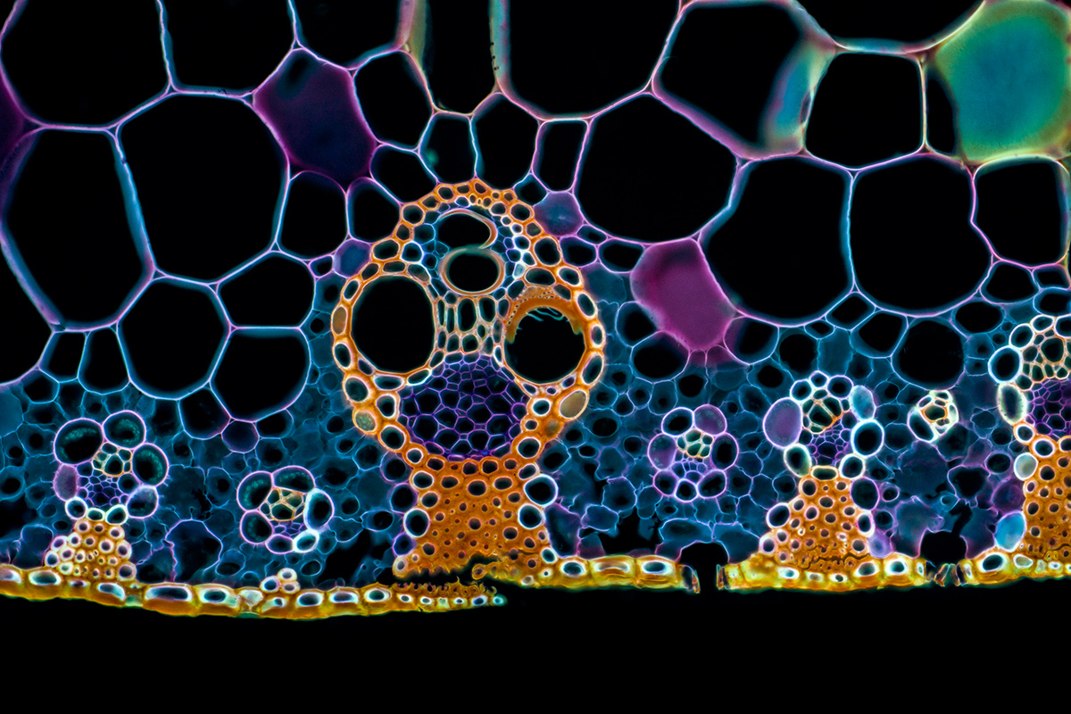
About the Micro-Wildlife Art
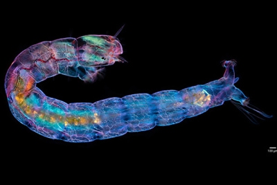
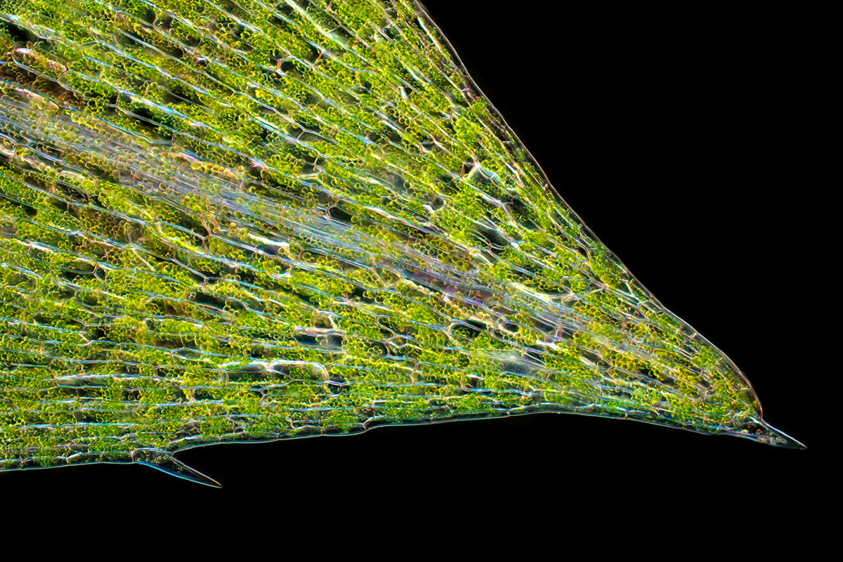
Karl carries out all of his imaging work from his home imaging laboratory. Throughout the year, he collects samples from various freshwater habitats and takes them back to the lab. The samples are poured into Petri dishes for inspection under a stereo microscope (Olympus SZX-16). Once a creature of interest is found, it is transferred, for a few moments, into a dish containing 70% alcohol in order to anesthetize it for imaging. The subject is then transferred to a slide and a coverslip mounted on top for imaging on the widefield microscope (Olympus BX51). The image of the Diptera larva was created for the 2022 Olympus Life Science calendar and to create it, the microscope was configured for compensated polarised dark field imaging. The compensation means that there is a wave plate inserted into the light path. The waveplate converts phase variations into different colours. Different wave plate thickness gives different colour palettes. When a subject is prepared and placed on the stage, Karl experiments with different lighting techniques to find which provides the most interesting and aesthetically pleasing result. The camera is rotated in order to compose the image and when doing montage imaging, in a way such as to minimise the number of stacks to be acquired. The microscope is equipped with the X Line range of Olympus objectives and the 10x/0.40 objective was used to create this image. The image is a focus stack montage. Images were acquired with the Sony A7RII, and images processed (aligned, stacked, and seamlessly blended by hand) in Adobe Photoshop.
Karl thrives to create imagery that are not only educationally enlightening, but are aesthetically stunning, to the degree that they draw people in for a closer look and to the extent that you would like to hang it on the walls of your home or office.
About the Chemical Art
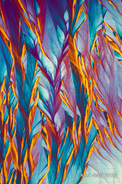
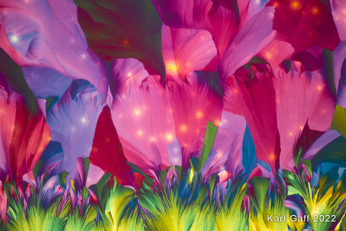
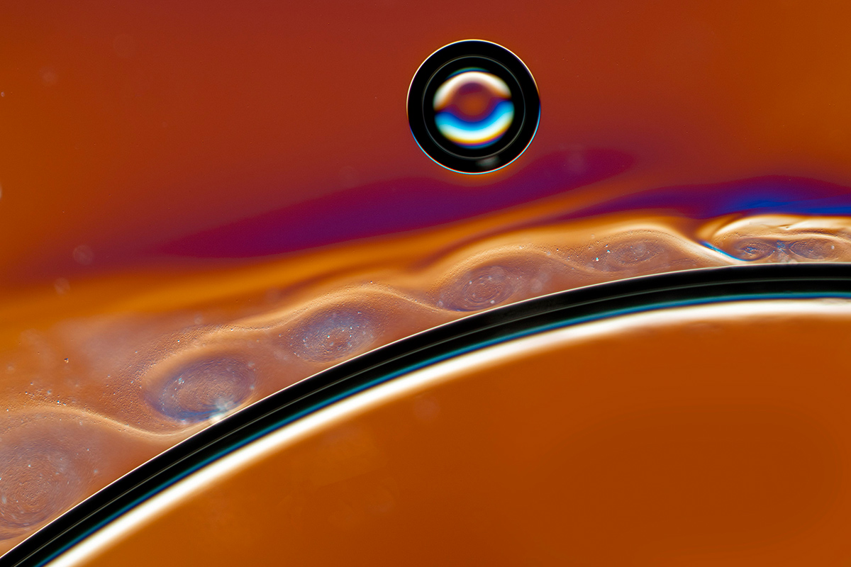
Karl thrives to create imagery that are not only educationally enlightening, but are aesthetically stunning, to the degree that they draw people in for a closer look and to the extent that you would like to hang it on the walls of your home or office. He carries out all of his imaging work from his home imaging laboratory. The photographic artworks presented here explore the mysterious worlds of chemicals. The journey into chemical space involves several stages. The first stage involves the formulation of the method for preparing a chemical and identifying the ingredients. Karl has kept a detailed recipe book of his findings and preparations for the past decade since beginning this photographic exploration. In the lab each day, he builds upon previous works by making small advancements to his methods and notes the results and observations. Karl explains that his work is a form of alchemy, which is defined as the process of taking something ordinary and turning into something extraordinary, sometimes in a way that cannot be explained. It is all about experimentation. The preparation involves mixing of particular chemicals in specific ratios and tweaking of those ratios. Although all steps are logged in his recipe book, sometimes he finds that it is not possible to reproduce a similar crystallographic pattern produced previously. This is likely because there are many aspects that influence the pattern formed, such as the presence of impurities and environmental factors such as variations in the working temperature and variations in the ambient humidity. However, this adds to the thrill because you never really know what to expect when you look through the eyepieces of the microscope!
Once a chemical solution is made, a droplet is smeared on a slide and allowed to crystallise before being transferred to the microscope stage for observation and photography. The artworks are not single photographs. They consist of numerous images stacked together into a single in-focus image. This is done because the sample is not entirely flat, it has a roughness to it, and because the microscope has a shallow depth of field, a number of images must be captured at different levels through the sample. Large panoramas can also be made that consist of focus-stacked montages, where a number of image stacks are captured over an area of the sample and seamlessly combined. This kind of photograph can then be printed to a very large size.
View more of Karl's work at https://www.kgaffphotography.com