Ultra Violet Laser Light - Ken Meats FBCA
© Ken Meats
This photo is of the ultra violet laser light source for an OMX microscope, taken by Ken Meats to accompany a news story about the first OMX scope in Canada, cutting edge technology at the time. Because it was to appear in a newspaper, the images had to be clear enough to demonstrate the device in black and white and in a low-res environment. This was one of two images used in the final article in The Globe and Mail.
"The photo was taken in a darkroom with a Nikon D200, Nikon AF-S DX Zoom Nikkor 17-70mm f/3.5-4.5 IF-ED, with the camera tripod mounted and the protective glass filter taken off the lens, as it eliminated the UV beams that can be seen in the image. Some plain glass protective filters can filter out UV light. A series of images was taken using a variety of exposures. This image was a 25 sec exposure at f/8. A very low power handheld fill flash was used to lighten the darker areas of the image."
"The difficulty in photographing this image was that there was no way to meter the lighting and the exposure was long and arrived at experimentally. Apart from the exposure time, photographing a UV light source is relatively simple with a modern DSLR. It took about two hours to do the whole shoot of which this photo was a part. The first part of the job was to figure out what I was looking at and establish how to shoot it."
Read more about this photographer
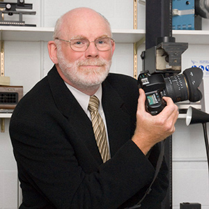
 Ken Meats was senior manager of Graphics and New Media at the Mount Sinai Hospital in Toronto, where he supported research at the Samuel Lunenfeld Research Institute. Part of his job was to help develop or refine photographic protocols for research projects. He currently works as a freelance medical photographer/designer, and has been developing his skills in wildlife photography. He is also a Registered Biological Photographer, a certification he earned through the BCA.
"I was interested in science and medicine all my life," he said. "I was also interested in photography and audio-visual and before a planned university degree I took courses in photography at a local community college. In my last couple of years of high school I became more interested in pursuing photography professionally and enrolled in a three-year photography program at Fanshawe College in London, Ontario."
During his first year as a full-time photography student, he discovered medical photography when his class toured a medical photography department at a local teaching hospital. "I realized that this field combined my interest in science and photography," he said. "With the department manager and medical photographer at the hospital and the program director at the college, I was able to develop a graduation portfolio with a medical emphasis. This led to my first job as a photo lab tech and to a long career that took me around the world."
Below, Meats shares insight into the field of medical photography and advice for other photographers interested in the field.
Describe your typical workday.
Working in a modern teaching hospital, there is no typical working day. Rather than describe my day as a manager, I'd prefer to describe a day in the department. In my department in addition to myself, we had a project coordinator, designers, one of whom was photographer, and a videographer. The videographer, designer/photographer or I did photography based on the type and volume of work. The photography could include a variety of events, portraits or studio setups for the Hospital Foundation, the communications department, or others, as well as clinical procedures in surgery, ER or elsewhere. Photographs are also taken for researchers of equipment or projects they are working on in the Research Institute. The video tech is also involved in providing video conferencing for rounds or in-service training.
What is the most used computer-editing tool in your workflow?
When working as a photographer, my most important tools are Adobe Photoshop Lightroom, Adobe Photoshop Extended and Adobe Bridge. The most difficult part of photography is keeping the images well catalogued. I find Lightroom and Bridge to be invaluable in maintaining the files.
As my work carries over into design and audio visual, I use most of the Adobe suite with InDesign being my primary design software and Adobe Audition the primary audio software.
What elements are important to you when you judge or critique your work or the work of other professional photographers?
The most important thing for a professional photographer is a sharp image. I think that all photographers have at times missed focus on a subject and it is one of the more embarrassing mistakes one makes. The other things, just as important for a great photograph, are the clarity of the area of interest and the composition of the whole image.
Do you have any advice for photographers interested in a career in biomedical/life sciences photography?
Learn the skills that other photographers don't need. In addition to a good grasp of audio and video, learn the terminology and basic physiology necessary to talk to the non-media professionals that you will be working with.
