
Uveitis - Paul Crompton FBCA
© Paul Crompton
This image, taken by Paul Crompton, is one of a series of photos of a case of post-operative uveitis, taken primarily for teaching purposes, although it also served as a record of the patient's condition for their case notes.
"The patient in this photo has developed an infection following an operation to replace his biological lens with an artificial one, called an Intra-Ocular lens or IOL. The lens is held in a capsule and the infection has caused the iris to adhere to the anterior surface of the capsule, leaving behind fragments of pigment."
"The photograph was produced on a Topcon photo-slit lamp fitted with a Nikon D2x. The slit lamp has a range of magnifications and this was taken at approximately 2.5x magnification. The camera was fitted with a flash (strobe) unit. There is no aperture so the exposure is controlled by varying the output of the flash unit. The human eye is a brilliant subject and the photo-slit lamp is a wonderful instrument for the photographer to use to capture the eye's complexities. You can control the height, width, direction, intensity and nature of the light to capture pathology in the different layers, textures and surfaces of the eye."
"The camera has very little depth of field (remember there is no aperture to stop down) so focusing is critical. You are also working with a living, breathing subject, at a high magnification, so patient management is really important. Making sure the patient is looking where you want them to look, remembering that their vision is compromised as well, that their eye is clearly open, but that they are reasonably comfortable and know what you are doing. You have to be at ease with your equipment, fully in control of it, because you have to be able to communicate with the patient at the same time as working the camera and lighting."
Read more about this photographer
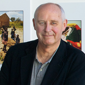 Paul Crompton began his career at Blackpool College of Art in the UK, where he specialized in advertising and commercial photography, but as he neared the end of his coursework, he felt that it wasn't quite the path he wanted to take.
"I had worked my way through college, some of the time in hospitals as a porter and cleaner and I really liked the atmosphere," he said. "There were also regular jobs coming up in medical photography so I applied to join a training scheme in Cardiff and have never really looked back from there. I did take a 10 year break from medical photography to teach, then came back to Cardiff as head of medical photography, just as digital was coming in. It was a great time to come back to the profession."
Below, Crompton shares insight into his interest in using light to capture the essence of a subject, and advice for photographers entering the field of medical photography.
Describe your typical workday.
As a manager my typical day is fairly desk-bound, although I do try to get behind the camera as much as I can. As for my team, their days are really varied. They could be photographing patients solidly, based in our ophthalmology, dental or dermatology units. The photographers working out of the main unit could be up on the wards, or in the operating theatres (OR in the States) photographing open-heart or orthopaedic surgery, which are two of our hospital specialties. We often get called to theatre for interesting and unusual cases, or if the surgeons are trying out a new technique. They may do that in the morning then be doing PR (public relations) work in the afternoon; anything from visiting 'stars' (Catherine Zeta Jones is one of our patrons) to portraits to 'grip-and-grins' for our communications department. The variety is one of the great things about the job but it can be stressful too.
What is the most used computer-editing tool in your workflow?
We use Adobe Photoshop as our main editing tool. It is integrated with our Digital Image Management system, which is the Fotoware range of products. This delivers images to our clinician's desktops through our hospital intranet. The workflow is really smooth, it has to be; we process about 20,000 patients a year.
What elements are important to you when you judge or critique your work or the work of other professional photographers?
What I am looking for in images is that thing I mentioned at the start, communication of an idea, a concept, some information (as in a clinical image) with the skills and craft of photography used to help, get the message across, preferably without drawing attention to itself. I am interested in seeing craft skills such as lighting, composition, framing, the use colour and tone, point of focus and depth of field and of course timing.
Do you have any advice for photographers interested in a career in medical photography?
Try to get some experience in a hospital or science environment. There is no substitute for it. It helps you to confirm that it is somewhere you want to work and it shows a prospective employer that you have some idea of how they work and that you are serious about working in the field. I am always interested in people who have wide photography skills and can demonstrate it in a portfolio. Photography is photography, whether it is fashion, portraiture, sports, documentary; the photographer's eye can be seen in all these.
I am as interested in what work students have done personally as much as what they have produced for college briefs. College work is fine but I want to see them express themselves through the medium. A couple of last thoughts; always have something to say about every picture in your portfolio and never say 'this is just a picture I took to', drop the 'just' bit, and finally, remember you are only as strong as your weakest image, if you can see something wrong in a picture, you can bet your life a prospective employer will!